Fig. 2.

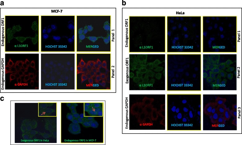
Immunofluorescence analysis reveals that α-human L1 ORF1p (RRM) can detect over expressed and endogenous ORF1p in its native conformation. a Panel 1: Endogenous ORF1p (green) detection in MCF-7 cells using α-human L1 ORF1p (RRM). Hochst (blue) was used to stain nuclear DNA. A merged image is shown (top right). Panel 2: Endogenous GAPDH protein served as a control of IHC technique. b Panel 1: Detection of endogenous ORF1p in HeLa cells (Left column).Nuclear DNA was stained with Hochst (middle column). Merged image shown in (top-right). Panel 2: Detection of exogenous ORF1 (green) in HeLa cells after transfecting pcDNA ORF1F [ORF1 green (left column), Hochst blue (middle column), merged (rightmost column)]. Panel 3: Endogenous GAPDH expression in HeLa cells. c Mostly cytoplasmic localization of ORF1p in MCF-7 (endogenous) and HeLa (exogenous) cells; however, some cells display nuclear localization of ORF1 protein (indicated by arrow)
