Figure 1.
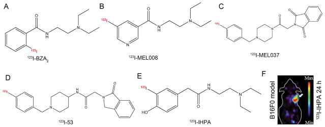
Representative melanin-targeting probes. Chemical structures of (A) 123I-BZA2, (B) 123I-MEL008, (C) 123I-MEL037, (D) 123I-53, and (E) 131I-IHPA. (F) MicroSPECT image of C57BL/6 mice bearing B16/F0 melanotic melanoma at 24 h postinjection of approximately 11.1 MBq of 123I-IHPA, the tumor is indicated by the white arrow. Adapted and modified with permission from references [34, 36, 41, 42, 44].

