Figure 2.
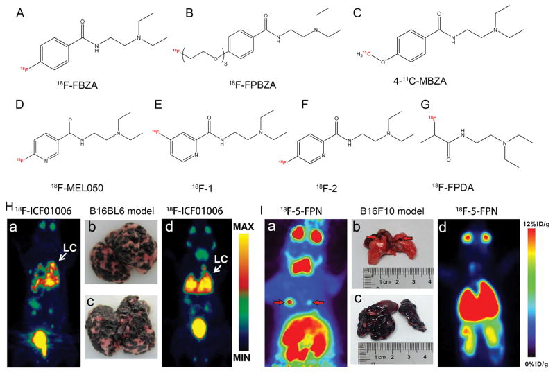
Representative melanin-targeting probes and in vivo study images. Chemical structures of (A) 18F-FBZA, (B) 18F-FPBZA, (C) 4-11C-MBZA, (D) 18F-MEL050, (E) 18F-1, (F) 18F-2, and (G) 18F-FPDA. (H) Whole-body maximum intensity projection (MIP) images of 18F-ICF01006 and corresponding lung photographs of B16/BL6 melanoma-bearing mice at the early stage (a, b) and late stage (c, d) of tumor development. (I) 18F-5-FPN PET images of two mice with lung metastases from melanoma. Note that this probe was able to detect both micrometastases (a, b) and wide spread lung metastases (c, d) from melanoma. Tumors are indicated by red arrows. Adapted and modified with permission from references [45–47, 49, 51, 52, 66, 67].

