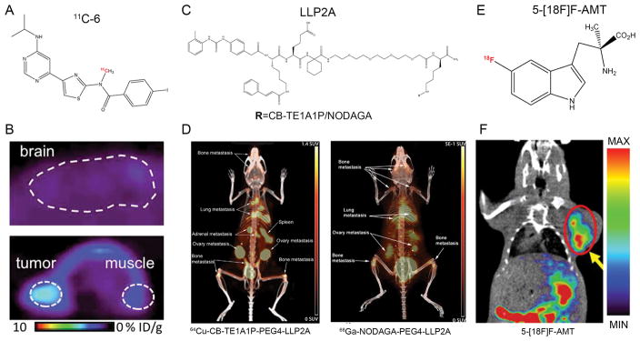
Figure 4.
Other PET probes for melanoma imaging. (A) Chemical structure of 11C-6 for imaging of mGlu1 receptor. (B) Representative PET images of mice bearing B16/F10 after injection of 11C-6. Upper: sagittal image of the brain. Lower: Axial image of tumor and muscle. (C) Chemical structures of NODAGA-PEG4-LLP2A and CB-TE1A1P-PEG4-LLP2A. (D) Small animal PET/CT imaging at 2 h after injection of the radiotracers (7.4 MBq). Both 64Cu-CB-TE1A1P-PEG4-LLP2A and 68Ga-NODAGA-PEG4-LLP2A were able to image melanoma lung metastases with high contrast and minimal lung background. (E) Chemical structure of 5-[18F]F-AMT. (F) Coronal PET/CT image of B16/F10 melanoma 30 min after injection of 5-[18F]F-AMT. Adapted and modified with permission from references [166, 172, 180].

