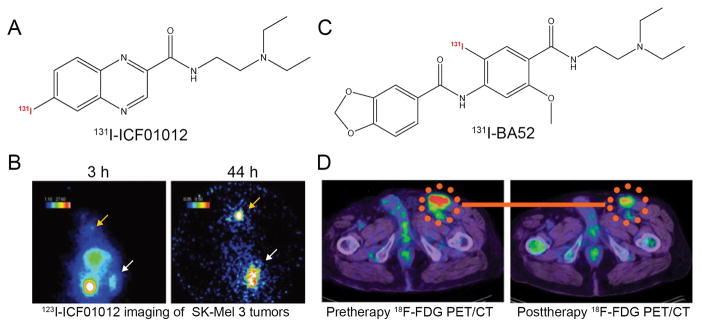
Figure 5.
Representative radionuclide-labeled therapeutic agents for melanoma. (A) Chemical structure of 131I-ICF01012. (B) Gamma-camera imaging of SK-Mel 3 melanoma-bearing mice after injection of 3.7 MBq 123I-ICF01012. A clear concentration of 123I-ICF01012 occurred 3 hours after radiotracer administration and a rapid elimination of 123I-ICF01012 from non-specific organs was observed at 44 h postinjection. Tumor and thyroid were indicated by white and orange arrows, respectively. (C) Chemical structure of 131I-BA52. (D) 18F-FDG PET/CT examinations in a melanoma patient before and after 131I-BA52 treatment. After treatment using 131I-BA52, post-therapeutic 18F-FDG PET/CT examination demonstrated that SUV of the inguinal lymph node metastasis decreased from 9.02 to 5.81. Adapted and modified with permission from references [221, 223].

