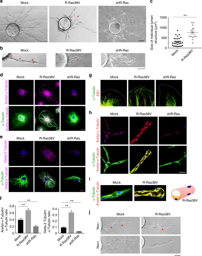
Fig. 1.
R-Ras is required for microtubule stabilization and EC lumenogenesis in vitro. a Endothelial sprouts in 3-D fibrin gel culture were analyzed at day 5. Arrow, anastomosed adjacent sprouts forming a continuous lumen. b Mock-transduced control EC culture in higher magnification showed incomplete lumen formation in the sprouts (arrows). Lumen formation (arrowhead) was significantly accelerated and enhanced in R-Ras38V-transduced ECs. R-Ras knockdown by shRNA (shR-Ras) blocked tubular morphogenesis and lumen formation. c Size of individual lumen structure. The hallow structures of >5 µm length were considered as developing lumens. The area size of the individual lumen structure was determined by morphometry analysis and plotted on the graph. Sprouts grown from 10 EC-coated beads in two culture wells were analyzed for each group. Small vacuoles of <5 µm in length were not included in the analysis. Spouts without lumen were not examined. No lumen was formed by R-Ras-silenced ECs. p < 1 × 10⁻⁵. d–i R-Ras stabilizes endothelial microtubules. Immunofluorescence of the total (green) and acetylated α-tubulin (d) or delta 2-tubulin (e) (magenta). f The ratio of post-translationally modified α-tubulin (acetylated α-tubulin or delta 2-tubulin) to the total α-tubulin was quantified from immunofluorescence staining of the cells. **p < 0.001. g Immunofluorescence of α-tubulin and microtubule end-binding protein (EB1) to indicate the (+) ends of microtubules. Thin gray lines indicate the outlines of the cell membrane. Lower magnification images available in Supplementary Fig. 4. h In-gel staining of endothelial sprouts for total (green) and acetylated (red) α-tubulin in 3-D culture (day 7) is shown by confocal sectional images. Yellow color indicates co-staining. i Higher magnification to show the pattern of extending microtubules. Acetylated microtubule fibers are depicted by yellow lines in the schematic representation of an R-Ras38V-expressing sprout in cross-section. j Nocodazole was added at 10 µM to 5-day-old culture of mock or R-Ras38V-transduced EC sprouts. Images of the sprouts were taken before (Noc−) and 1.5 h after (Noc+) nocodazole treatment. Arrows indicate two discontinuous lumens in a control sprout and a complete uninterrupted lumen in an R-Ras38V+ sprout. Scale bars, 75 µm (a), 50 µm (b, h, j), 20 µm (d, e)

