Fig. 5.
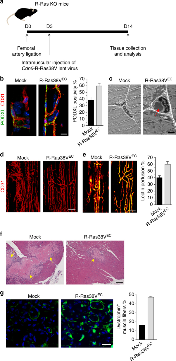
RRAS gene delivery to ECs rescues vessel lumenogenesis and muscle reperfusion. a In vivo transduction and treatment schedule. The lentivirus carrying pLenti6/Cdh5-R-Ras38V expression vector was injected into GC muscles 3 days after ligation. GC muscle were analyzed at day 14. b Immunofluorescence of CD31 and PODXL to identify lumenized vessels in GC muscles after the lentivirus injection for EC-specific expression of R-Ras38V (R-Ras38VEC). PODXL positivity % (PODXL+CD31+ area/CD31+ area × 100) was determined to assess the fraction of lumenized vessel area. c Transmission electron microscopy of the GC muscles confirmed the increase in vessel lumenization upon R-Ras38VEC transduction. Arrow, a circulating erythrocyte found in the vessel lumen indicating normal lumen formation. d CD31 staining of whole-mounted GC muscle fascicles. e Analysis of vessel perfusion in whole-mounted GC muscle fascicles. Yellow color indicates lectin perfused vessels. f H&E staining of GC muscle sections. Arrows, necrotic areas. g Dystrophin immunostaining (green) of GC muscle cross-sections to quantify functional muscle fibers. The number of dystrophin+ muscle fibers/total muscle fibers (%) was determined in non-necrotic area. p < 10−4, n = 5. Scale bars, 25 µm (b), 3 µm (c), 100 µm (d), 50 µm (e), 150 µm (f)

