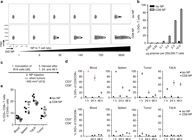
Fig. 2.
CD8a-targeting nanoparticles bind to T cell in vitro and in vivo. a CD8a-targeting nanoparticles (loaded with DiD), but not isotype control nanoparticles, bind to the surface of CD8+ T cells isolated from the spleen within 30 min of incubation, as assessed by flow cytometry. b Quantification of DiD-positive T cells; data representative for more than 4 experiments. c Timeline of in vivo binding experiment. d Quantification of DiD-positive, CD3+/CD8+ and CD3+/CD8− T cells 1, 24, and 48 h after the nanoparticles were injected intravenously; n = 3 for 1 and 48 h, n = 6 for 24 h, mean ± s.e.m.; (red open circle) Anti-CD8a antibody for flow cytometry staining could not bind due to steric hindrance with CD8a-targeting nanoparticles. e Quantification of CD3/CD8+ T cells in blood, spleen, tumor-draining lymph node (TdLN), and tumor 24 h after the nanoparticles were injected intravenously; n = 6

