Fig. 5.
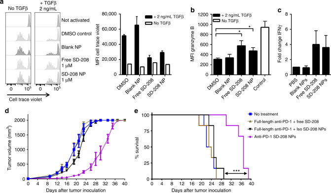
Targeted delivery of a TGFβR1 inhibitor (SD-208) to PD-1-expressing cells delays tumor growth and extends survival. a Proliferation of CD8+ T cells following activation with anti-CD3/CD28 beads (1:2 bead to T cell ratio) for 72 h in the presence or absence of TGFβ1 (2 ng mL−1) and treatment with 1 μM SD-208 as free compound or as nanoparticle formulation; quantification of geometric mean of cell trace violet is provided in the right panel, n = 3, mean ± s.d. b Intracellular granzyme B expression was assessed by flow cytometry, n = 3, mean ± s.d. (*p < 0.05, one-way ANOVA with Tukey’s post hoc test). c Fold change of interferon-γ (IFNγ) was measured by ELISA, n = 4, mean ± s.e.m. d, e C57BL/6 were inoculated subcutaneously with MC38 cells. Five days later, nanoparticles or free drugs were administered intravenously every other day up to a total of 10 injections. The dose was 20 μg of anti-PD-1 and 40 μg of SD-208. d Tumor volume and e animal survival were monitored to assess efficacy, n = 6, mean ± s.e.m.; (***p < 0.001, Mantel-Cox test)

