Fig. 3.
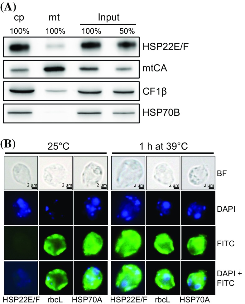
Localization of HSP22E/F to the chloroplast. a Subcellular localization of HSP22E/F by immunoblotting. 10 or 3 µg protein (depending on the antiserum used) from whole cells (input), chloroplasts (cp) and mitochondria (mt) isolated from strain cw15-302 exposed to 39 °C for 60 min were separated by SDS-PAGE and immunodetected with antisera against HSP22F, mitochondrial carboanhydrase (mtCA), extrinsic thylakoid membrane protein CF1β, and stromal HSP70B. b Microscopy images taken from cells of strain cw15-325 that were kept at 25 °C or exposed to 39 °C for 60 min. Shown are from top to bottom: bright field (BF) images, DAPI staining, immunofluorescence (FITC), and the merge of DAPI and FITC. Antisera used for immunofluorescence were against HSP22F, stromal RbcL, and cytosolic HSP70A

