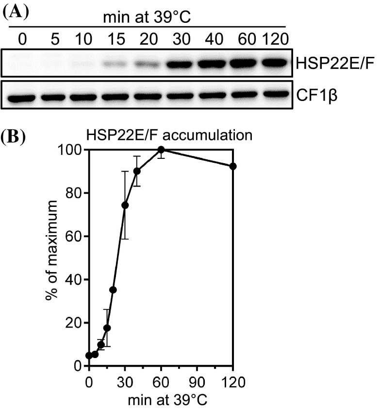
Fig. 4.
Accumulation of HSP22E/F during heat stress. a Immunoblot analysis of HSP22E/F accumulation during heat stress. cw15-302 cells grown at 25 °C were exposed to 39 °C for 120 min. Total proteins corresponding to 0.25 µg chlorophyll from cells harvested at the time points given were separated by SDS-PAGE and analyzed by immunoblotting using antisera against HSP22F and against CF1β as loading control. b Quantification of HSP22E/F signal intensities from (a). Signals were normalized to the maximal HSP22E/F levels reached during the time course. Error bars represent SD, n = 2

