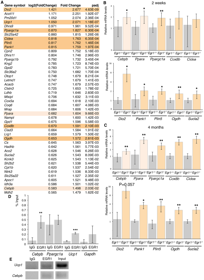
Figure 3.
Transcriptomic analysis of subcutaneous inguinal adipose tissue of postnatal Egr1 −/− versus Egr1 +/+ mice shows upregulation of beige adipocyte markers. (A) List of the first 45 upregulated genes in 6 inguinal subcutaneous fat pads of 3 Egr1 −/− versus 3 Egr1 +/+ 2-week-old mice. (B,C) RT-qPCR analysis of the expression levels for generic adipocyte differentiation markers Cebpb, Ppara, beige adipocyte differentiation marker, Ppargc1a, Cox8b, Cidea, Dio2, Pank1, Plin5, Ogdh and Sucla2 in SC-WAT of 2-week-old (B) and 4-month-old (C) Egr1 −/− mice compared to Egr1 +/+ mice. For each gene, the mRNA levels of control (Egr1 +/+) SC-WAT were normalized to 1. Graphs show means ± standard deviations of 5 samples from 2-week-old Egr1 +/+ mice and Egr1 −/− mice, 6 samples from 4-month-old Egr1 +/+ mice and 5 samples from Egr1 −/− mice. The relative mRNA levels were calculated using the 2−ΔΔCt method. The p-values were obtained using the Mann-Withney test. Asterisks indicate the p-values *p < 0.05, **p < 0.01. (D) Chromatin Immunoprecipitation (ChIP) assays were performed from 60 fat pads of wild type 2-week-old mice with antibodies against EGR1 or IgG2 as irrelevant antibody in three independent biological experiments. ChIP products were analyzed by RT-q-PCR (N = 2). Primers targeting the proximal promoter regions of Cebpb and Ucp1 revealed the recruitment of EGR1 in the vicinity of these sequences, while primers targeting the proximal promoter regions of Ppargc1a and Gapdh (negative controls) did not show any immunoprecipitation with EGR1 antibody compared to IgG2 antibody. Results were represented as percentage of the input. Error bars showed standard deviations. The p-values were obtained using the Mann-Withney test. Asterisks indicate the p-values, **p < 0.01, ***p < 0.001. (E) ChIP-qPCR samples were loaded on agarose gel and confirmed a specific amplification of Cebpb and Ucp1 promoter regions after chromatin immunoprecipitation using EGR1 antibody. No DNA was immunoprecipitated by irrelevant IgG antibody. Input chromatin was diluted 2 times for Ucp1 qPCR and 4 times for Cebpb qPCR.

