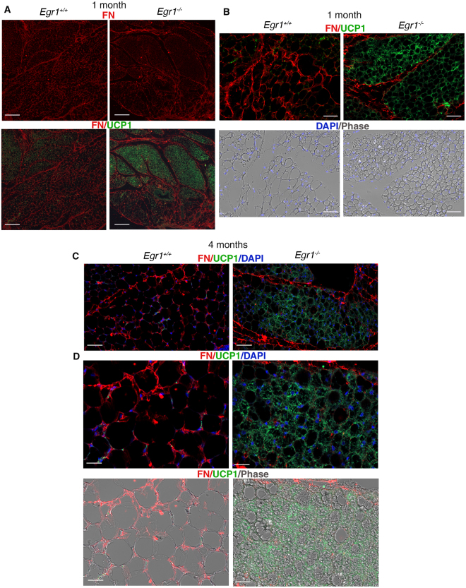
Figure 5.
Fibronectin localization in SC-WATs of Egr1 +/+ and Egr1 −/− mice. SC-WATs of 1-month-old (A,B) and 4-months-old (C,D) Egr1 +/+ and Egr1 −/− mice were sectioned transversely and immuno-stained with Fibronectin FN (red) and UCP1 (green) antibodies. Nuclei were visualized with DAPI (blue). Individual channel or merged channels are indicated in panels. (A) Low magnifications show that FN (red) is less expressed in UCP1-positive areas (green) compared to UCP1-negative areas in 1-month-old Egr1 +/+ and Egr1 −/− mice. Scale bars, 200 µm. (B) High magnifications show that FN is absent around UCP1 + beige adipoctytes in 1-month-old Egr1 −/− compared to Egr1 +/+ mice, while being present around white adipoctyes in Egr1 +/+ mice. Scale bars, 50 µm. (C,D) At 4 months of age, FN is also absent around UCP1 + beige adipoctyes of SC-WATs from Egr1 −/−, while being present around white adipocytes in Egr1 +/+ mice. Scale bars, (C) 50 µm (D) 25 µm. (B–D) FN surrounds the regions of beige adipocytes.

