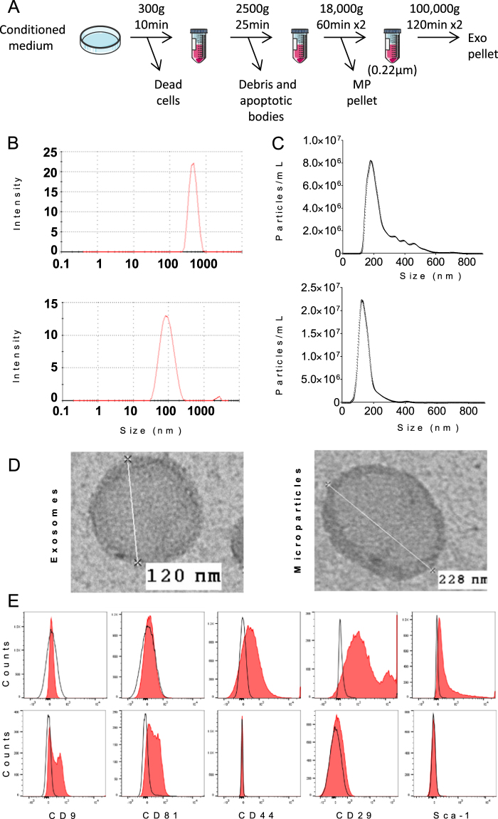
Figure 1.
Isolation and characterization of extracellular vesicles isolated from murine BM-MSCs. (A) Experimental protocol for isolation of microparticles (MPs) and exosomes (Exos) using differential ultracentrifugation. (B) Size of MPs (up) and Exos (down) detected in 200 µL by Dynamic Light Scattering analysis (C) Number and size of MPs (up) and Exos (down) detected in 1 mL (corresponding to 1 µg EV equivalent proteins) by Nano Tracking Analysis. (D) Representative images of Exos and MPs by transmission electron microscopy. (E) Expression of BM-MSC membrane markers (Sca-1, CD44, CD29) and of exosomal markers (CD9, CD81) on MPs (top) and Exos (bottom) isolated from naïve BM-MSCs as analysed by flow cytometry.

