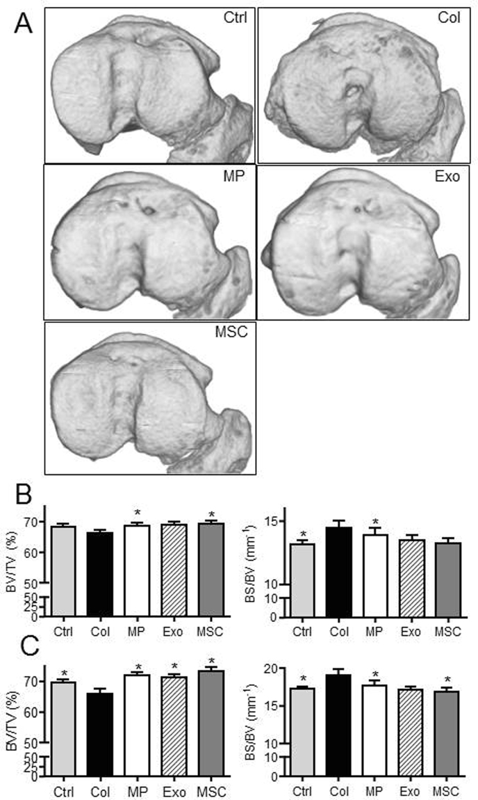
Figure 5.
BM-MSC-derived MPs and Exos protected mice from osteoarthritic damages in the collagenase-induced OA model. (A) Representative 3D reconstructed images of sub-chondral bone surface in tibias after µCT analysis. Images from control mice (Ctrl), collagenase-treated mice (Col) and Col mice that received intra-articular injection of 500 ng MPs or 250 ng Exos or 2.5 × 10⁵ BM-MSCs. (B) Histomorphometric analysis of 3D images of epiphyseal bone: Bone volume/tissue volume (BV/TV) and bone surface/bone volume (BS/BV) parameters (n = 15). (C) Histomorphometric analysis of sub-chondral bone. *: p < 0.05 as compared to Col group.

