Figure 1.
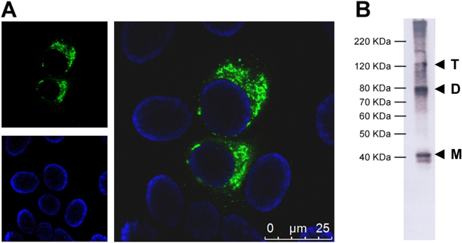
Characterization of 3A8-C12 MAb reactivity. (A) Indirect immunofluorescence of YFV/17DD-infected Huh7.5 cells (confocal microscopy) stained with the 3A8-C12 monoclonal antibody plus anti-mouse IgG conjugated with AlexaFluor 488. DNA was stained using DAPI; (B) Western blotting analysis of rNS1 resolved by 15% SDS-PAGE. The membrane was stained with the 3A8-C12 monoclonal antibody, followed by anti-mouse IgG conjugated with alkaline phosphatase. Solid arrows indicate oligomeric forms of YFV rNS1: (M) monomers, (D) dimers and (T) trimers.

