
Figure 1.
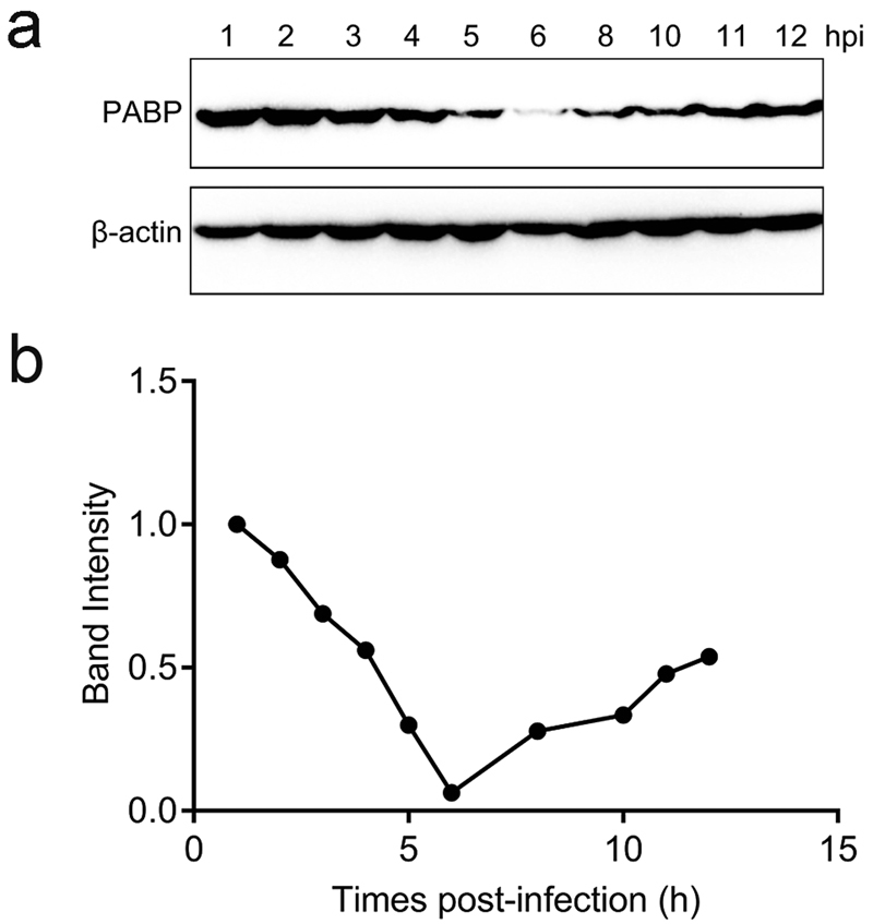
Change in cellular poly(A)-binding protein (PABP) during the duck hepatitis A virus (DHAV) infection of duck embryo fibroblast (DEF) cells. (a) Western blotting analysis was performed to detect the protein expression levels of PABP, with β-actin as the loading control. (b) Band intensity of PABP in the DHAV infection. The band intensities representing PABP protein expression levels were quantitated using the β-actin control bands as a reference (for each time point) using the Image J software.