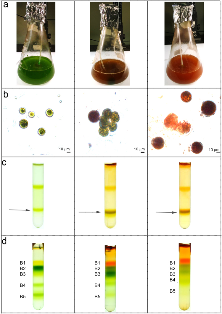
Figure 1.
Cell cultivation, membranes and pigment binding complexes isolation. Panel a: H. pluvialis cultures grown under 3 different stress conditions. Green: 50 µmol m−2s−1 for 7 days; Orange: 400 µmol m−2s−1 for 3 days; Red: 400 µmol m−2s−1 for 6 days. Panel b: microscope observation of cells grown as in Panel A. Panel c: isolation of plastid membranes from G, O and R cells. Purified membranes are indicated by the arrow. Panel d: Sucrose gradient ultracentrifugation separation of pigment binding complexes from plastid membranes solubilized in β-DM 1%.

