Figure 1.
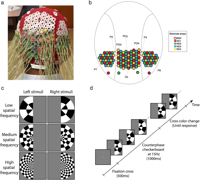
Experimental design. (a) Modified EEG head cap with high-density electrode configuration. (b) Map of electrode configuration, showing the SND layout and subset of electrodes designated for each ND array. Some standard electrode locations are marked for reference. (c) Stimuli and design of the experiment: stimuli were low, medium or high spatial frequency checkerboards presented in the left or right visual field. (d) An example trial. A central gray fixation cross was presented for 500 ms, then the checkerboard stimulus counter-phased at 15 Hz. After 1000 ms, the cross became red or green, at which time participants discriminated its color and responded via a key-press.

