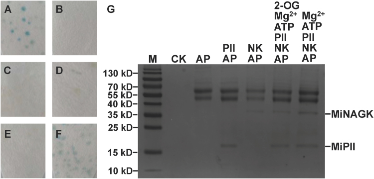
Figure 2.
Protein-protein interaction between MiPII and MiNAGK as detected by yeast two-hybrid (from A through F) and co-immunoprecipitation (G) assays. Images A through E show yeast co-transformed with a pair of either pGBKT7-53 and pGADT7-T as a positive control (A) or pGBKT7-Lam and pGADT7-T as a negative control (B). From C through F iamges, yeast co-transformed with either pGBKT7-glnB only (C), pGBKT7-glnB and pGADT7-accB1 (D), pGBKT7-glnB and pGADT7-accB2 (E), or pGADT7-argB and pGBKT7-glnB (F). In Image G, 6 μL, 3 μL, and 5 μL of 1 M Mg2+, ATP, and 2-OG as marked above the panel, respectively, were added to 1 mL solution containing 200 μg and 400 μg recombinant MiPII and MiNAGK, respectively. The presence of MiNAGK bound to the Sepharose beads was subsequently analyzed by SDS-PAGE (15% acrylamide) and Coomassie blue R-250 staining. Lane M, protein molecular marker. AP, anti-MiPII polyclonal antibody. CK, only Sepharose as a control. NK, MiNAGK.

