Figure 8.
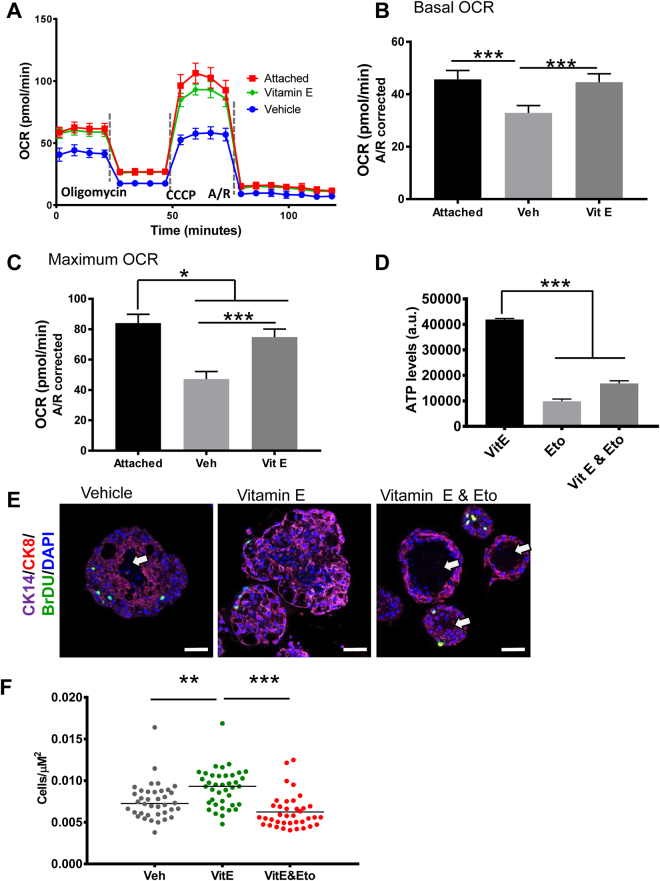
Vitamin E restores ATP levels in detached cells by stimulating fatty acid oxidation leading to organoid luminal filling. (A) OCAR analysis of RWPE-1 cells grown in adherent and non-adherent conditions and treated with the SELECT supplements for 24 h, n = 15. (B) Basal OCAR was calculated by subtracting non-mitochondrial OCAR from the basal OCAR measurements. (C) Maximal OCAR was calculated by subtracting non-mitochondrial OCAR from the OCAR measurements after the injection of CCCP. (D) Detached RWPE-1 cells were treated with vehicle, vitamin E or vitamin E and an FAO inhibitor, Etomoxir (Eto, 25 μM) for 24 h; ATP measurement showed that FAO inhibition abrogated the ATP rescue by Vitamin E. (E) Immunostained sections of RWPE-1 organoids from (D), showed that the Vitamin E treated organoids had filled lumens while those co-treated with Vitamin E and Etomoxir or vehicle had hollow lumens (arrows). (F) Cell density of organoids from (E) measured by dividing the number of total cells per organoid by its area showed that the Vitamin E organoids had the highest cell densities while those co-treated with Vitamin E and Etomoxir had the lowest cell densities. Scale bars represent 100 μm. Asterisks represent statistical significance (One way ANOVA with Tukey’s correction for multiple comparisons). **p ≤ 0.05, **p ≤ 0.01, ***p ≤ 0.0001, error bars represent SD.

