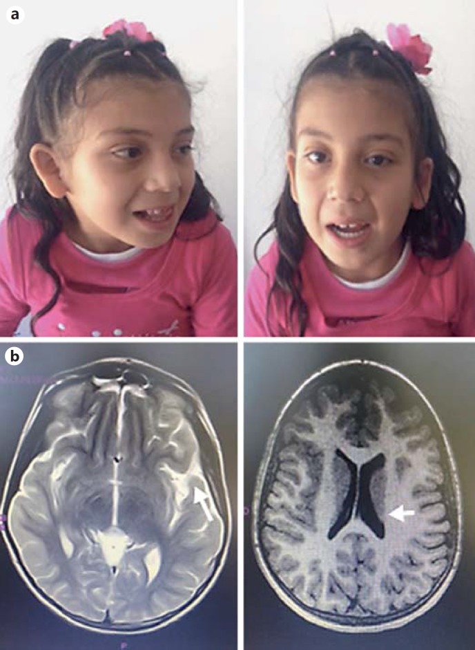
Fig. 1.
a Patient at the age of 5 years. Facial features showing midfacial hypoplasia, hypertelorism, micrognathia, epicanthic fold, prominent teeth, and upslanting palpebral fissures. b Brain MRI showing frontal and temporal cortical atrophy, with loss of posterior ventricular white matter (arrows).

