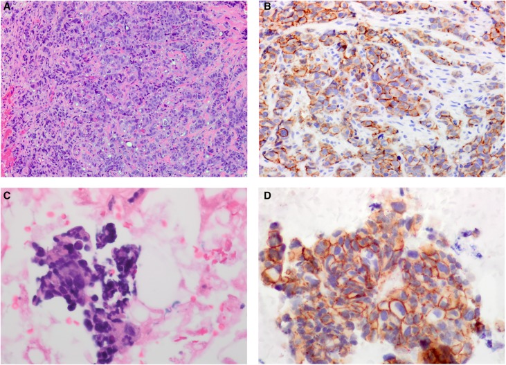
Figure 1.
Histopathology of the MLAV. (A) The biopsy of the vulvar mass shows a poorly differentiated tumor composed of nests and cords of pleomorphic tumor cells (20× magnification). (B) The HER2 immunostain on the biopsy of the vulvar mass is equivocal, compatible with score 2+ based on predominantly incomplete, weak and moderate membrane staining within >10% of tumor cells (20× magnification). (C) The fine needle aspirate of the supraclavicular lymph node shows clusters of pleomorphic tumor cells consistent with metastatic mammary-like adenocarcinoma (40× magnification). (D) The HER2 immunostain of the supraclavicular lymph node shows tumor cells with complete, intense membrane staining in >10% of tumor cells compatible with score 3+ (40× magnification).

