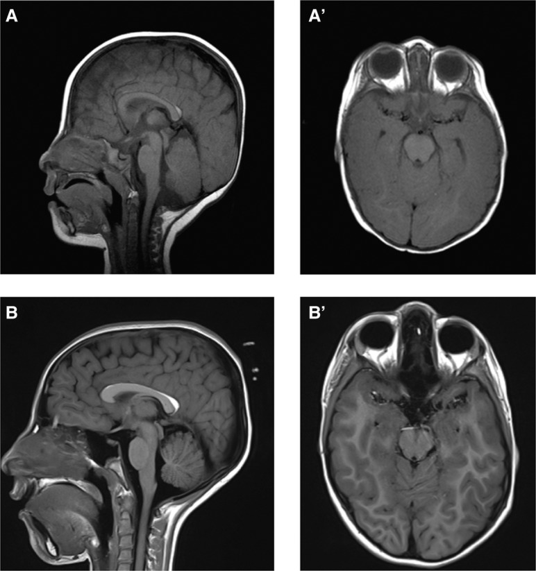
Figure 3.

Brain imaging data of individual with EBF3 variant. (A,B) Sagittal and (A′,B′) axial images of Individual 4 at 9 mo (A,A′) and 9 yr (B,B′) of age show a mild hypoplasia of the cerebellar vermis, mildly dysplastic corpus callosum, and small pericallosal lipoma without evidence of significant progression.
