Fig. 4.

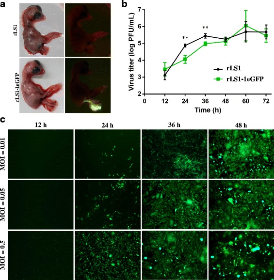
Characterization of rLS1-1eGFP virus. a SPF chicken embryos infected with rLS1 or rLS1-1eGFP. The embryos were inoculated at 9 days of age and incubated for 3 days. The eGFP expression was clearly observed at the umbilical cord of the embryos. b Comparison of the growth kinetics of rLS1 and rLS1-1eGFP. DF-1 cells were infected with an MOI of 0.05 and supernatant was collected in 12 h intervals post-infection and tittered by plaque assay. The data were taken from three independent experiments. Statistical significance (p < 0.01) was denoted with two asterisks. c Correlation between eGFP expression and different MOIs and times of infection. DF-1 cells were infected with rLS1-1eGFP at indicated MOIs and eGFP expression observed at 12, 24, 36 and 48 h.p.i
