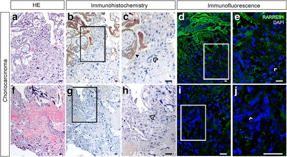
Fig. 3.

RARRES1 expression in choriocarcinoma sections. Sections of choriocarcinomas ((a-e) intramolare choriocarcinoma and (f-j) pure choriocarcinoma; n = 2 representative patients) were stained with anti-RARRES1 antibody by immunohistochemistry (a-c and f-h) or immunofluorescence (d, e, i and j). RARRES1 positive staining is indicated in brown (a-c and f-h) or green (d, e, i and j), nuclei are blue. RARRES1 positive cells are marked by black arrow heads; white arrow heads show RARRES1 negative cells; (c, e, h and j) are magnifications of the sections in (b, d, g and i) marked by squares, respectively. The bar equals 100 μm
