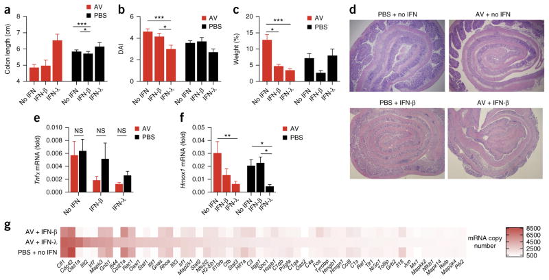
Figure 8.
IFN-λ displays a superior ability in suppressing DSS-induced colitis in the absence of enteric viruses, by diminishing oxidative stress. (a–d) Colitis development in wild-type mice treated intragastrically with a ‘cocktail’ of AV drugs or PBS for 10 d before the administration of 2.5% DSS in the drinking water, as well as no interferon (no IFN), mouse recombinant IFN-λ to which polyethylene glycol was attached (IFN-λ) or recombinant IFN-β (IFN-β) (200 U per gram body weight per day of either), evaluated as colon length (a), disease activity (b), weight at day 7, relative to initial weight (c), and histology (d). (e,f) qPCR analysis of Tnf in the total colon (e) and of Hmox1 in intestinal epithelial cells (measuring oxidative damage) (f) in mice as in a–d. (g) NanoString analysis of gene expression (nCounter Mouse Inflammation v2 panel; genes that generated >500 counts per treatment) in mRNA (100 ng) purified from total colon extracts of mice as in a–d. *P < 0.05, **P < 0.01 and ***P < 0.001 (two-way ANOVA.). Data are from one of three independent experiments (mean + s.e.m. of five mice per group in a–c,e,f).

