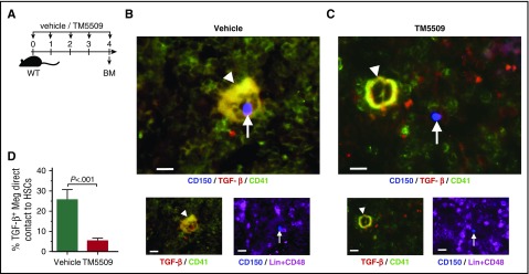
Figure 3.
PAI-1 regulates HSCs localization in the BM niche. (A) Schema for immunofluorescence analysis. (B-C) Representative pictures of the BM cavity of vehicle- or TM5509-treated mice. BM sections were stained with anti-CD150 (blue), anti-CD41 (green), anti-TGF-β (red), and anti-CD48 and -lineage markers (purple) antibodies. Arrowheads indicate TGF-β–expressing megakaryocytes. Arrows indicate Lin−CD48−CD150+ HSCs. Bars represent 100 μm. (D) Percentages of TGF-β–expressing megakaryocytes in close contact to HSCs. More than 100 cells in random fields on a slide were counted for 3 independent experiments. Means ± SD are shown in each bar graph. Meg, megakaryocyte.

