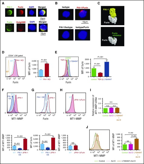
Figure 6.
iPAI-1 modulates MT1-MMP expression through the regulation of Furin. (A) Representative immunofluorescence microscopic images of PAI-1, Furin, and trans-Golgi complex in LSK CD34− cells. (B) D-PLA imaging shows the specific interaction between iPAI-1 and Furin in LSK CD34− cells. As a negative control for D-PLA, slides were treated with either the combination of anti-PAI-1 Ab and mouse IgG or anti-Furin Ab and rabbit IgG followed by the D-PLA secondary Abs. No fluorescent foci were detected by this treatment. (C) The docking simulations between Furin (white) and the active form of PAI-1 (yellow) show the tightly bound PAI-1 covering the active site of Furin, which prevents other substrates from approaching the active site (top). Compare with a structure of Furin (white) with a Furin inhibitor58 (green) bound at the active site of Furin (bottom). (D) Representative flow cytometric profiles and MFI (n = 6) for Furin expression in LSKCD34− cells isolated from WT or PAI-1 KO mice. (E) Representative flow cytometric profiles and MFI (n = 6) for Furin expression in LSK cells treated in vitro with either TGF-β or LY364947. (F-H) Representative flow cytometric profiles and MFI (n = 6) for MT1-MMP (F and H) and Furin (G) expression in PAI-1-overexpressed (OE), PAI-1-deleted (ΔPAI-1), or PAI-1/Furin double-deleted (ΔPAI-1/ΔFurin) hematopoietic cell lines. (D-H) Means ± SD are shown. (I-J) Relative messenger RNA (I) and representative flow cytometric profile (J) for MT1-MMP expression in LSK cells treated with LY364947 in the presence of actinomycin D in vitro. Bar graphs represent means ± SD (n = 5) of MT1-MMP expression. DAPI, 4',6-diamidino-2-phenylindole.

