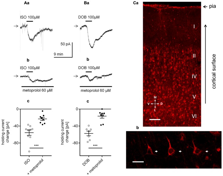
FIGURE 6.
Effect of β1-adrenergic receptor stimulation on the holding current in layer V mPFC pyramidal neurons. (A) Inward currents evoked by bath application of the non-selective β-adrenergic receptor agonist isoproterenol alone (ISO, 100 μM, a) and isoproterenol (ISO, 100 μM) in the presence of the selective β1-adrenergic receptor blocker metoprolol (60 μM in the bath, b). Amplitude of the holding current change evoked by 100 μM isoproterenol alone (ISO) and 100 μM isoproterenol in the presence of 60 μM metoprolol (+ metoprolol, c). (B) Inward currents evoked by bath application of the selective β1-adrenergic receptor agonist dobutamine alone (DOB, 100 μM, a) and dobutamine (DOB, 100 μM) in the presence of the selective β1-adrenergic receptor blocker metoprolol (60 μM in the bath, b). Amplitude of the holding current change evoked by 100 μM dobutamine alone (DOB) and 100 μM dobutamine in the presence of 60 μM metoprolol (+ metoprolol, c); ∗∗∗p < 0.001. (C) Immunofluorescent staining of β1-adrenergic receptor protein in the rat mPFC. The signal is localized to neurons in different cortical layers (a). Layer V at higher magnification with a pyramidal neuron showing immunofluorescent signal within its soma (arrows) and apical dendrites (b). Scale bars (a) 100 μm, (b) 25 μm. M, medial; L, lateral; D, dorsal; V, ventral.

