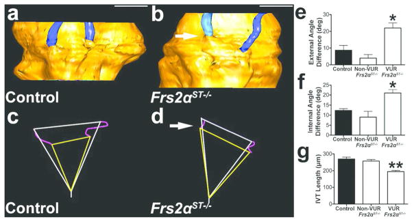
Figure 5. Refluxing ureters in P1 Frs2αST−/− mice have improper ureteral insertion and shortened intravesicular tunnel lengths.
(a–b) Representative 3D reconstruction reveal similar bladder insertion points of both control ureters (a, blue) into the bladder (yellow), but a high and lateral insertion of the right Frs2αST−/− ureter (arrow) into the bladder. (c–d) Triangles connecting the external ureter (white) and internal (yellow) insertion points and the bladder neck (from panels a and b) illustrate similar insertion site angles in the control, indicating relatively parallel levels of insertion (c); however, the high and lateral refluxing Frs2αST−/− ureter (arrow) leads to triangles with very different insertion point angles (d). (e–f) Graphs confirm that compared to control and non-refluxing Frs2αST−/− mice, Frs2αST−/− mice with unilateral reflux have much larger differences in external (e) and internal (f) insertion site angles. (g) Graph revealing that mean intravesicular tunnel length (IVT) (visualized as purple lines in panels c and d) is significantly shorter in refluxing Frs2αST−/− ureters versus control and non-refluxing Frs2αST−/− ureters. (n) = 3. Values = Mean + SD. One-way ANOVA = *p<0.05, **p<0.01. Scale bar = 500μm.

