
Fig. 8.
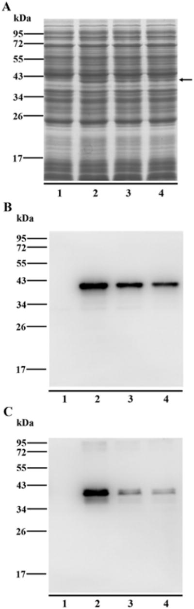
Western blotting analysis of the reactivity of anti-trout IgT1 mAb to other IgT subclasses. The CH2–CH4 regions of trout IgT1, IgT2 and IgT3 were recombinantly expressed in E.coli. Supernatants of the cell lysates (containing the soluble recombinant proteins) were subjected to SDS-PAGE under reducing condition for coomassie blue staining (A) and under both reducing (B) and non-reducing (C) conditions for immunoblotting with mouse anti-trout IgT1 mAb. The total proteins loaded on SDS-PAGE were 15 μg for gel staining and 5 μg for immunoblotting. The expressed recombinant proteins were directed by a black arrow. Lane 1: blank control (trout Igτ1 without IPTG induction); Lane 2: trout Igτ1; Lane 3: trout Igτ2; Lane 4: trout Igτ3.