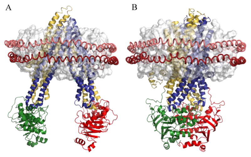
Figure 1.

P-gp in inward-facing and outward-facing conformations. (A) Crystal structure of apo murine P-gp (Protein Data Bank entry 4Q9H) and (B) illustrative homology model of human P-gp (see Materials and Methods) manually docked with a discoidal HDL model. Colored segments are transmembrane domains 1 (yellow) and 2 (blue) and nucleotide binding domains 1 (green) and 2 (red). The OPM database10 was used for the spatial arrangement of P-gp with respect to the lipid bilayer (white). The red helical peptides around the lipid bilayer represent the MSP protein of the nanodisc.
