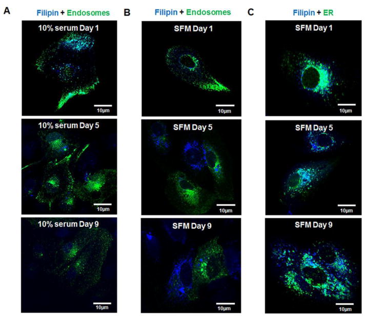
Figure 2. Confocal images showing changes in localization of UC cholesterol in cell organelles during serum deprivation.
A: Filipin (UC) and Endosome staining of cells in 10% medium from day 1–9. B: Filipin (UC) and Endosome staining of cells in SFM from day 1–9. C: Filipin (UC) and Endoplasmic Reticulum (ER) staining of cells in SFM from day 1–9.

