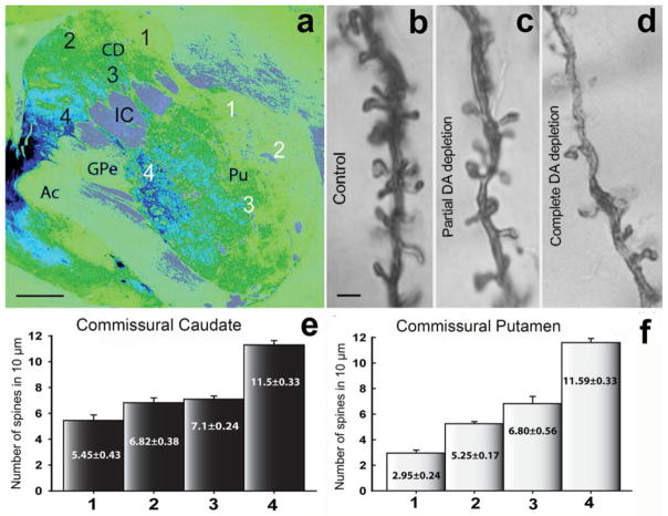
Fig. 2. Tyrosine hydroxylase (TH) immunoreactivity (a) and dendritic spine density in the striatum of partially dopamine depleted MPTP-treated monkeys (b–f).
a: Pseudo-colored image (NIH ImageJ program) of a TH-immunostained section from the monkey commissural striatum. The numbers (1–4) in the caudate (black numbers) and putamen (white numbers) indicate the areas from where the Golgi-impregnated neurons were selected for the dendritic spine analysis showed in e and f. The lowest (complete DA loss) and highest (no DA loss) levels of TH-immunostaining correspond to the numbers 1 and 4, respectively. b–d: Golgi impregnated dendrites (20–30 μm from the soma) from monkey striatal SPNs in control (no DA depletion; 10–12 spines per μm) (b), partial DA depletion (c), and complete DA depletion (4–6 spines per μm) (d). e, f: Histograms showing the dendritic spine density of Golgi-impregnated neurons from commissural striatal areas with different degrees of dopamine depletion. The values in the bars represent the mean of the spine density (mean±SEM) per 10 μm of dendritic length (primary dendrite) from neurons (10 neurons per area) from the corresponding striatal areas indicated by number (1 to 4) and color (black or white) in a. The Abbreviations: Ac: anterior commissure; CD: caudate; IC: internal capsule; Pu: putamen; GPe: Globus pallidus external segment. Scale bars in a: 1 mm, and in b (applies to c and d): 1 μm.

