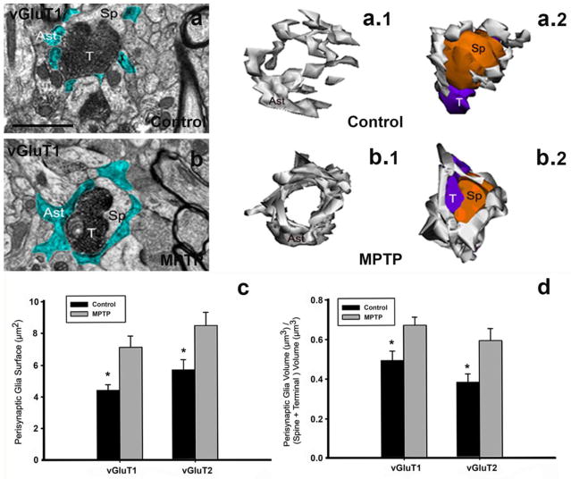
Fig. 4. Perisynaptic astrocytes in striatal axo-spine glutamatergic synapses of control and MPTP-treated monkeys.
a, b: Examples of single electron micrographs (EM) images of perisynaptic astrocytic processes (Ast) in control (a) and MPTP-treated monkey (b). Glial processes have been pseudocolored in blue (a, b). a.1, a.2, b.1, b.2: Three–dimensional reconstruction of axo-spine synapses formed by a vGluT1-positive terminal (T) and a dendritic spine (Sp) in the striatum of control (a.1, a.2) and MPTP-treated parkinsonian (b.1, b.2) monkey. While axo-spinous interfaces in control (a, a.1, a.2) are only partially surrounded by astroglial processes (Ast), those synapses are almost completely wrapped by enlarged astrocyte (Ast) processes in MPTP-animals (b, b.1, b.2). c, d: Quantitative analysis of the perisynaptic glia in striatal glutamatergic axo-spine synapses. c: Comparison of the surface area (mean±SEM) of perisynaptic glia associated with axo-spine synapses formed vGluT1- and vGluT2-immunopositive terminals in control and MPTP-treated monkeys. The surface of the perisynaptic glia is significantly larger (*, t-test; SigmaPlot) in MPTP-parkinsonian monkeys than in control (p=0.017 for vGluT1, and p=0.06 for vGluT2). d: Comparison of the volume of the perisynaptic glia over the total volume of the spine and the vGluT1- or vGluT2-immunoreactive terminal. This ratio is significantly increased in axo-spines synapses from MPTP-treated animals compared with control (*, t-test, p=0.049 for vGluT1 and p=0.028 for vGluT2, SigmaPlot). No significant difference is found between the volume of perisynaptic glia in axo-spine synapses formed for vGluT1- or vGluT2-positive terminals. Number of animals=3 controls and 3 MPTP-treated monkeys. Total number of reconstructed spines=32 (8 per group). Scale bar in a (applies to b): 1μm.

