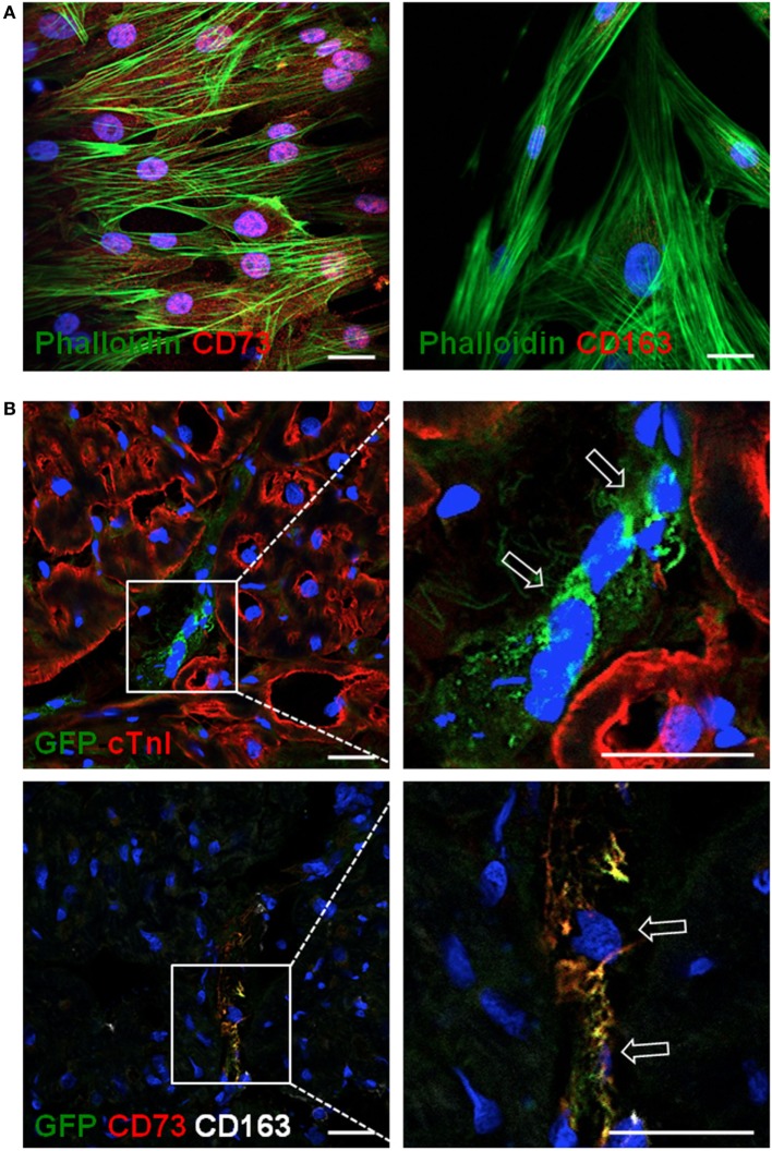
Figure 6.
Allogeneic cATMSCs migrated to the infarcted myocardium after graft implantation in swine. Representative confocal microscope images showing (A) porcine cATMSCs in in vitro culture or (B) sections within the infarcted myocardium. (A) Porcine cATMSCs are positive for CD73 (left, red) and negative for CD163 (right, red). (B) Presence of GFP+ porcine cATMSCs (empty arrows) in post-infarcted myocardium, also positive for CD73 but negative for CD163 (lower panels). Cell morphology, cardiac muscle, and cell nuclei are also counterstained using Atto 488-phalloidin, anti-cTnI Ab, and DAPI, respectively. Scale bars = 50 µm. cATMSCs, cardiac adipose tissue-derived MSCs; DAPI, 4′,6-diamidino-2-phenylindole dihydrochloride.

