Fig. 6.
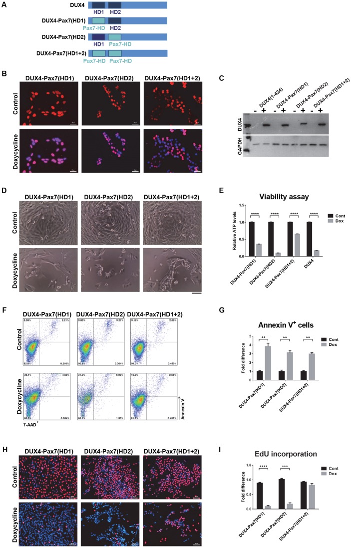
Toxicity of DUX4-Pax7 homeodomain substitutions. (A) DUX4-Pax7 homeodomain substitutions. In the construct DUX4-Pax7(HD1) the first DUX4 homeodomain was substituted, in DUX4-Pax7(HD2) the second, and in DUX4-Pax7(HD1+2) both homeodomains were substituted with the mouse Pax7 homeodomain. (B) Immunostaining revealed nuclear localization of hybrid proteins (red). Cells were induced for 20 h with 250 ng ml−1 doxycycline. (C) Western blotting with RD247c antibody shows the protein expression from the hybrid constructs. (D) Cell morphology after 48 h induction with 250 ng ml−1 doxycycline. Obvious cell death was observed with induction of all three hybrid constructs. (E) ATP assay for cell viability after 48 h of induction with 500 ng ml−1 doxycycline (n=8). (F) FACS analyses of cells induced with 500 ng ml−1 doxycycline for 18 h and stained with Annexin V/7-AAD. (G) Percentage of Annexin V-positive cells after 18 h induction with 500 ng ml−1 doxycycline (n=4). (H) Representative images of cells labeled with EdU. Cells were induced for 12 h with 250 ng ml−1 doxycycline and labeled for an additional 12 h with EdU. EdU-labeled cells are stained with red and nuclei were counterstained with Hoechst 33342 (blue). (I) EdU incorporation in cells presented in H (n=6). All data are presented as fold difference compared with control (uninduced) cells; mean±s.e.m., one-way ANOVA. **P<0.01, ***P<0.001, ****P<0.0001. Scale bars: 50 µm (B,H), 100 µm. (D)

