Fig. 6.
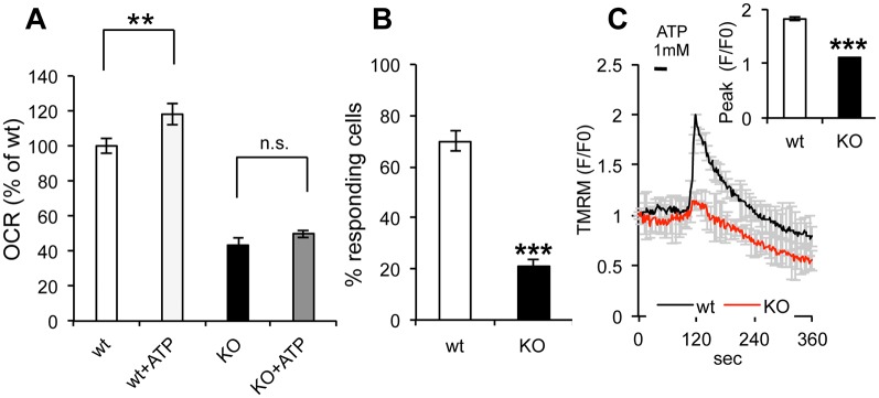
ER-mitochondria bioenergetics is coupled through Ca2+ signaling. (A) OCR measured in uncoupled mitochondria (SF 1 µM) of wt and sAC KO cells in the presence or absence of ATP (n=3). (B) Percentage of responding wt and sAC KO cells loaded with fluo4 and stimulated with ATP 1 mM. (C) TMRM average intensity of wt and sAC KO MEFs upon ATP 1 mM stimulation and loaded with fluo4. Insert: peak average intensity of fluo4 in wt and sAC KO MEFs upon ATP stimulation (n=30−55, in 3 independent experiments). Data is expressed as mean±s.e.m. in indicated number of different biological replicates (**P<0.01, ***P<0.001, Student's t-test).

