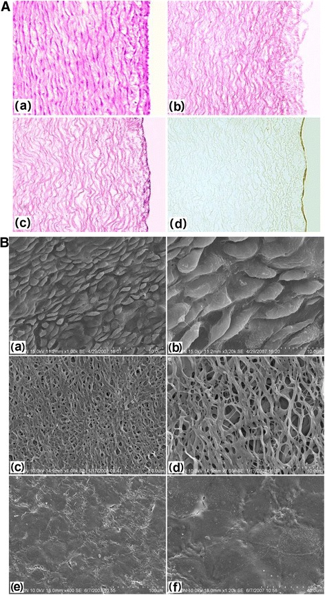
Fig. 3.

Histological and immunohistochemical staining and scanning electron microscopy of specimens. A: a, histological staining of fetal pig aorta; b: histological staining of decellularized aortae of fetal pigs (DAFPs); c: histological staining of tissue-engineered vascular grafts with an EC layer; d: immunohistochemical staining of ECs of vascular grafts. B: a: fetal pig aortic EC layer at low magnification; b: fetal pig aortic EC layer at high magnification; c: the inner elastic membrane of DAFPs at low magnification; d: the inner elastic membrane of DAFPs at high magnification; e: the EC layer of tissue-engineered vascular grafts at low magnification; f: the EC layer of tissue-engineered vascular grafts at high magnification
