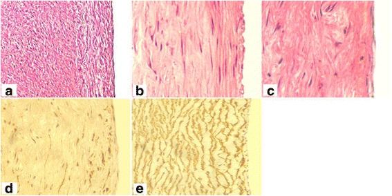
Fig. 6.

Histological staining of tissue-engineered vascular grafts after implantation. a, b, and c, Hematoxylin–eosin staining of tissue-engineered vascular grafts at 6 months post-implantation; d smooth muscle actin (SMA) immunohistochemical staining of vascular grafts; e SMA immunohistochemical staining of fetal pig aorta as a positive control
