Fig. 7.

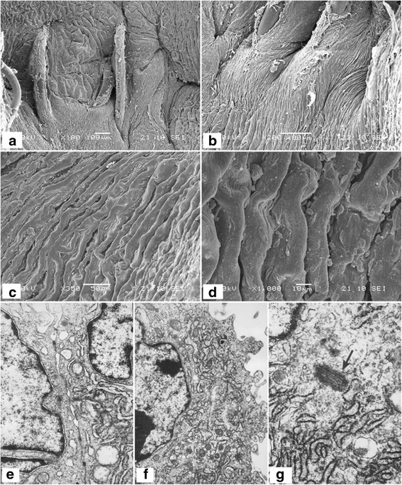
Electron microscopy of tissue-engineered vascular grafts at 6 months post-implantation. a–d, the inner surface of vascular grafts by scanning electron microscopy; e, close connections between ECs were identified by transmission electron microscopy (TEM). f, the cell cytoplasm was rich in active organelles identified by TEM. g specific structures of ECs as seen at high magnification by TEM
