Fig. 1.
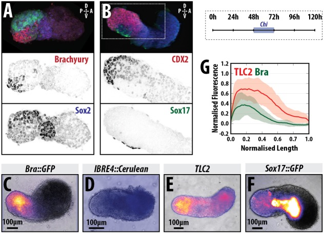
Axial organisation of gastruloids. (A,B) Sox1::GFP (A) and Nodal::YFP reporter (B) gastruloids pulsed with Chi (48-72 h AA) and stained with Hoechst and anti-GFP with either (A) T/Bra (red) and Sox2 (blue), or (B) Cdx2 (red) and Sox17 (green) at 120 h AA; Hoechst is not shown in A; staining is representative of at least three replicate experiments; 3D projections are displayed. (C-F) Gastruloids formed from T/Bra::GFP (C), BMP (IBRE4::Cerulean; D), Wnt/β-catenin (TLC2; E) and Sox17::GFP (F) reporter lines following a 48-72 h Chi pulse. (G) Quantification of reporter expression for the TLC2 (red) and T/Bra::GFP (green) gastruloids in a posterior-to-anterior direction. Stimulation results in activation of the TLC2 reporter with highest expression at the posterior pole. Schematic for the stimulation regime is shown in the top-right corner. Scale bars: 100 μm in C-F.

