Fig. 3.

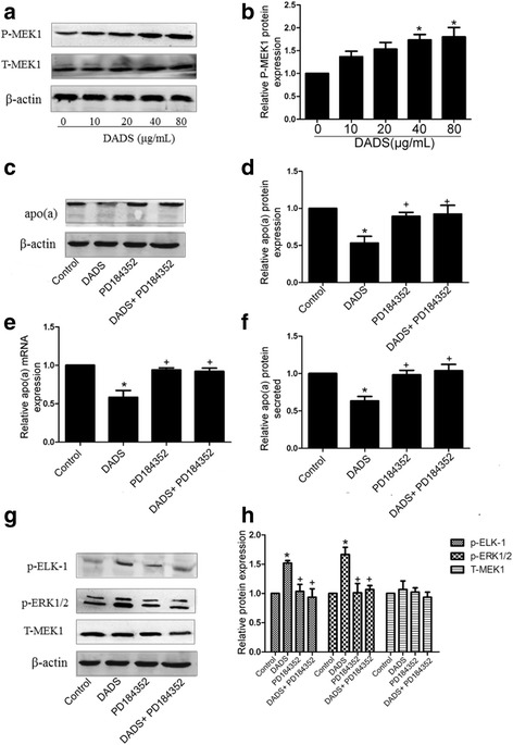
Involvement of MEK1 activation by DADS in apo(a) expression suppression. a, b DADS increased phosphorylated MEK1 in a dose-dependent manner, as determined through Western blot analysis. HepG2 cells were treated with specific inhibitors of MEK1(PD184352) for 6 h and with DADS (40 μg/mL) for 24 h. c, d Western blot results and densitometric quantification of apo(a) expression in whole-cell lysates from all groups. e The mRNA levels of apo(a) were determined using RT-qPCR. f The levels of secreted apo(a) were determined by ELISA. g, h Western blot results and densitometric quantification of phosphorylated ERK1/2 and phosphorylated ELK1 levels in whole-cell lysates from all groups. Each experiment was performed in triplicate, and the results are expressed as mean ± SD, *p < 0.05 versus control; p < 0.05 versus DADS. MEK1 inhibitors were used for PD184352
