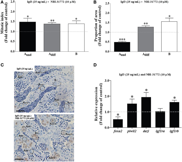
Figure 5.
Effect of 25 ng/mL Igf3 in the presence of an IGF-binding protein inhibitor on spermatogonial proliferation and proportion of area after 3 days of primary testis tissue culture. (A) Mitotic indices of type Aund, type Adiff, and type B spermatogonia in the presence of Igf3 alone (25 ng/mL) (dotted line; control condition) or in combination with 10 µM NBI-31772 (bars) (n = 6). (B) Proportion of section surface area occupied by cysts containing type Aund, type Adiff, or type B spermatogonia in the presence of Igf3 alone (25 ng/mL) (dotted line; control condition) or in combination with 10 µM NBI-31772 (bars) (n = 6). (C) Immunocytochemical detection of BrdU in sections of zebrafish testis incubated with 25 ng/mL alone (upper panel; control condition) or in combination with 10 µM NBI-31772 (lower panel) for 3 days showing BrdU positive (+) and negative (−) Aund, Adiff, and B spermatogonia. Bars, 25 µm. (D) Gene expression analysis in adult zebrafish testis after 3 days of tissue culture in the presence of Igf3 (25 ng/mL) (represented by a dotted line) or in combination with 10 µM NBI-31772 (bars) (n = 7). Results are presented as fold changes with respect to the control group (25 ng/mL Igf3). Asterisks indicate significant differences (*P < 0.05; **P < 0.01; ***P < 0.001) between groups.

