
Figure 3. STAT3 represses Rev-erbα transcription by binding directly to its promoter.
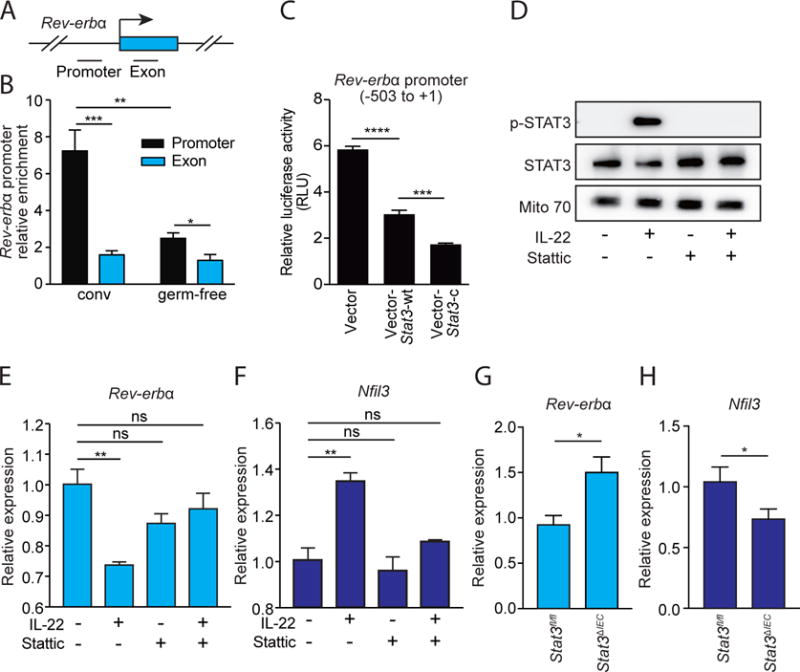
(A) Schematic of the Rev-erbα gene promoter. (B) ChIP analysis of intestinal epithelial cells from conventional (conv) or germ-free mice using immunoglobulin G (IgG) or anti-STAT3 antibody. Precipitated fragments of the Rev-erbα promoter or control exon were detected by qRT-PCR. (C) Luciferase reporter assay. A 504 bp fragment of Rev-erbα promoter was fused to a firefly luciferase reporter. HEK-293T cells were transfected with reporters and either empty vector, a wild-type STAT3-encoding vector (Stat3-wt), or a dominant active STAT3-encoding vector (Stat3-c). (D) Western-blot of total STAT3, phosphorylated STAT3 (p-STAT3) in small intestinal organoids treated with IL-22 and/or the STAT3 inhibitor Stattic. Mito 70 is the loading control. (E,F) qRT-PCR analysis of Rev-erbα (E) and Nfil3 (F) expression in small intestinal organoids treated with IL-22 and/or Stattic. (G,H) qRT-PCR analysis of epithelial Rev-erbα (G) and Nfil3 (H) expression in Stat3fl/fl and Stat3ΔIEC mice at ZT4. N=3–8 samples per group. Means±SEM are plotted; statistics were performed with Student’s t-test or one-way ANOVA. *p<0.05; **p<0.01; ***p<0.001; ****p<0.0001; ns, not significant.