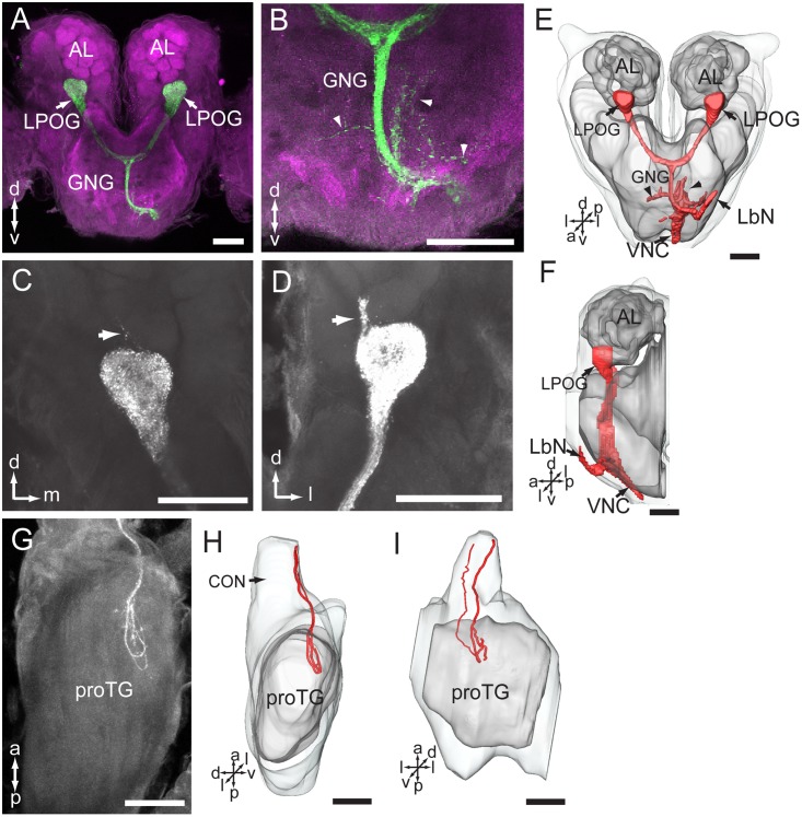
FIGURE 6.
Central pathway of sensory neurons located in the labial palp organ (LPO). (A) Confocal image showing the main target region of the LPO sensory neurons (green), i.e., the LPO glomerulus (LPOG) in each antennal lobe (AL; magenta). (B) Enlarged image showing the sensory processes in the gnathal ganglion (GNG; arrowheads). (C,D) Confocal images showing terminals of stained axons innervating the entire LPOG, with a few processes outside of LPOG (arrows). (E,F) Three-dimensional reconstructions of the sensory pathway shown in (A,B). (E) In frontal view, (F) in lateral view. (G) Confocal image showing axon projections in the prothoracic ganglion (TG). (H,I) Three-dimensional reconstructions of the axon terminals shown in (G). (H) In lateral view, (I) in ventral view. CON, connective; LbN, labial nerve; VNC, ventral nerve cord. a, anterior; d, dorsal; l, lateral; p, posterior; v, ventral. Scale bars, 100 μm.

