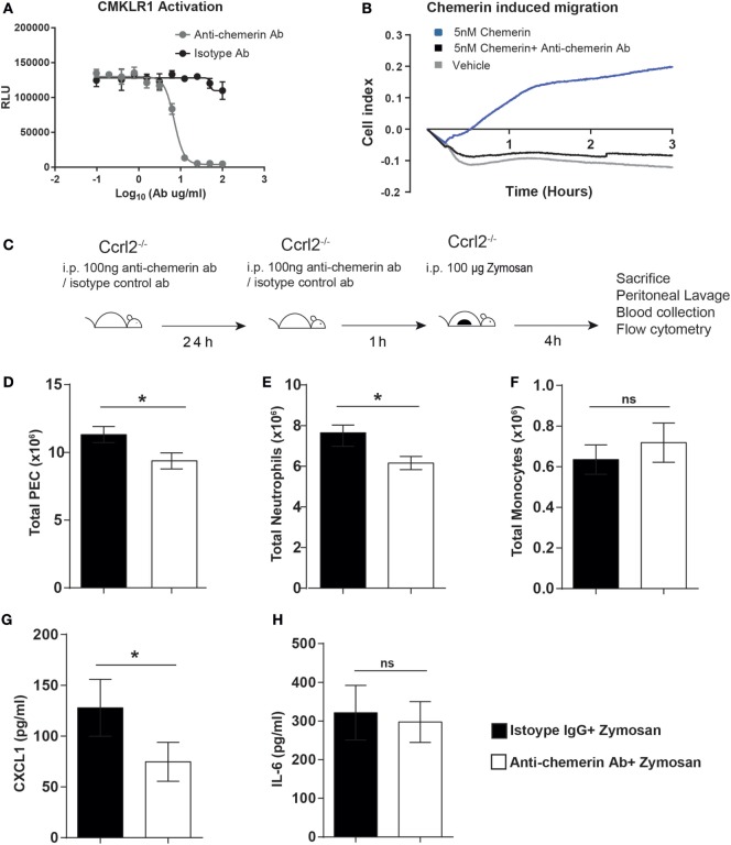
Figure 5.
Treatment with an anti-chemerin blocking antibody attenuated the exaggerated inflammatory responses observed in Ccrl2−/− mice. (A) CHO-K1 cells stably transfected with murine Cmklr1 were plated out in a 96-well plate for 48 h before stimulation. Cells were challenged with indicated concentrations of anti-chemerin antibody or isotype control before challenge with 20 nM murine chemerin. Cmklr1 activity was assessed by quantification of β-arrestin recruitment to Cmklr1 as measured by luminescence. RLU, relative light units. Error bars = SD of three technical replicates of one experiment. (B) Representative real-time chemotaxis trace of biogel-elicited peritoneal exudate cells (PECs). 8- to 10-week-old male C57BL/6J mice were injected i.p. with 2% Bio-gel (polyacrylamide beads) and sacrificed 4 days later. Peritoneal cavities were lavaged with ice-cold PBS supplemented with 2 mM EDTA. Cells were pretreated with vehicle or anti-chemerin antibody (ab) for 45 min. A gradient of 5 nM of the 5 nM chemerin was allowed to form, and chemotaxis was measured of 4 × 10⁵ cells (400,000 cells/well) for 3 h. (C) Schematic of in vivo experimental design. (D–G) 8- to 10-week-old male Ccrl2−/− mice were pretreated with 100 ng of anti-chemerin blocking antibody or isotype control IgG antibody i.p. for 24 h before challenge with zymosan for 4 h. Animals were sacrificed, and peritoneal cavities were lavaged with ice-cold PBS supplemented with 2 mM EDTA. Cells were quantified using counting beads, and cell populations were analysed using flow cytometry (D) Total cells. (E) Total neutrophils. (F). Total monocytes. Levels of CXCL1 (G) and IL-6 (H) in the peritoneum of indicated groups were quantified by ELISA. Mean ± SEM. n = 2–8 mice/group. Statistical significance was assessed using a Student’s unpaired t-test. *P ≤ 0.05, **P ≤ 0.01.

