
Fig. 3.
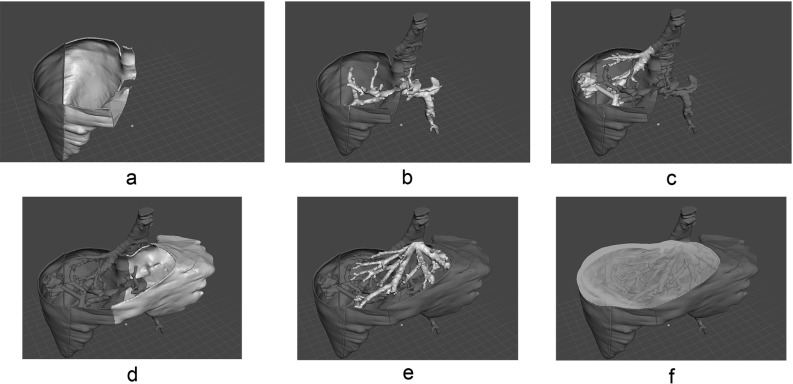
Assembly simulation. All of the parts in the 3D modeling software are shown in a common view. The goal is to determine the order of bonding with cyanoacrylate adhesive. In this study, the bonding order was as follows: a the two liver parenchyma parts; b the portal vein; c the inferior vena cava connected to the right hepatic vein and the tumor; d the third parenchyma part; e the left and middle hepatic vein; and f the fourth and final liver parenchyma part. This exact order was replicated during the assembly of the actual physical model