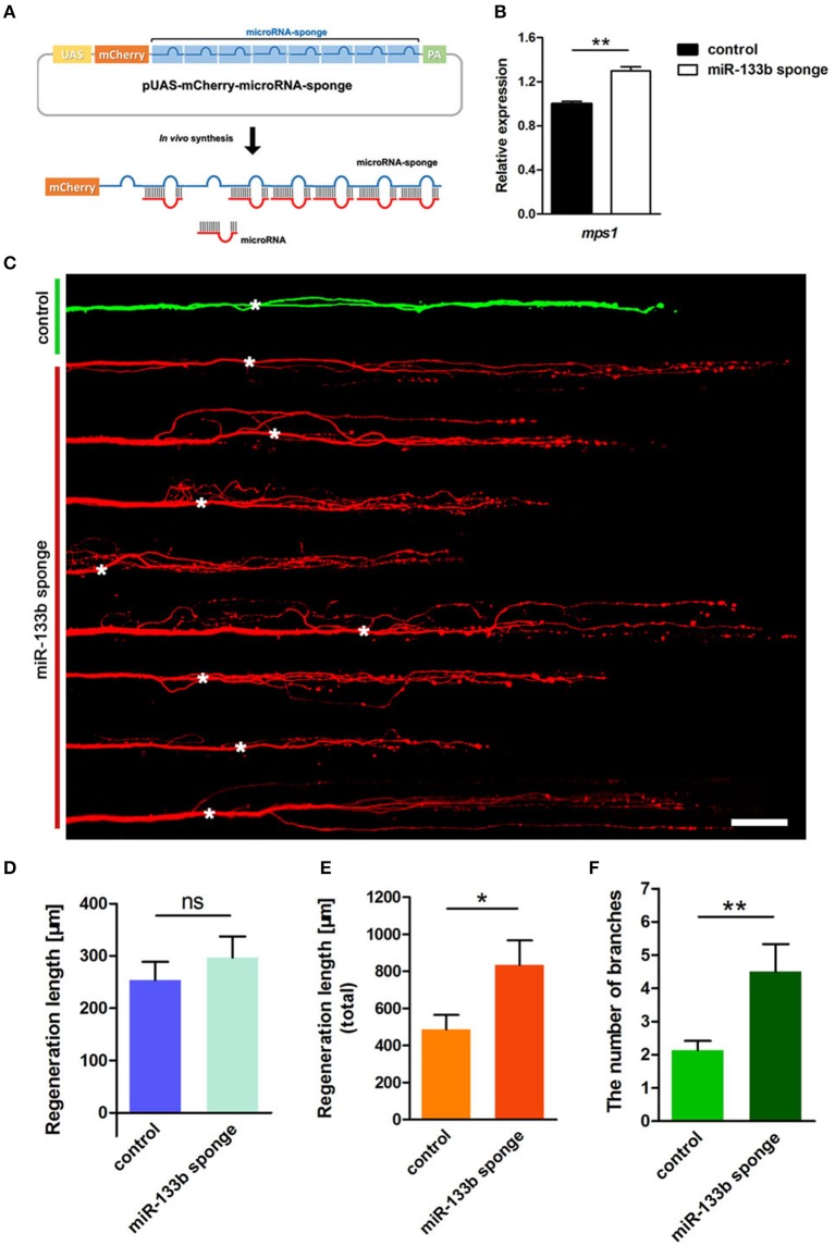
Figure 2.
Knockdown of miR-133b by expressing miR-133b sponge facilitates M-cell regeneration. (A) Design of miRNA sponges. The construction of miRNA sponges was manipulated by inserting multiple microRNA binding sites in the 3′-UTR of the mcherry reporter gene. Plasmids express only mCherry served as control vector. (B) Quantitative RT-PCR analysis exhibited an increase in mps1 mRNA expression in 10 hpf zebrafish embryos by miR-133b sponge expression in vivo. (C) Confocal imaging of M-cell at 2 dpa. White asterisk: ablation point. Scale bar: 50μm. (D) Regeneration length at 2 dpa. Student's two-tailed t-test, P = 0.4300. (E) Total regeneration length at 2 dpa. Student's two-tailed t-test, P = 0.0194. (F) The number of branches at 2 dpa. Non-parametric tests, P = 0.0047. *P < 0.05, **P < 0.001. Error bars represent S.E.M.

